SCIENTISTS MAY HAVE FOUND A WAY TO REPAIR DAMAGED EYES
A September 2026 study repaired defects in patient-derived retinal models. It offers a route toward treatment for RP11, not proof of restored human sight.
An experimental gene therapy has restored important functions in laboratory-grown retinal cells carrying an inherited disease. The advance is real, but its meaning is narrower than a cure for damaged eyes: no patient regained sight in this experiment. The work concerns retinitis pigmentosa type 11, or RP11, and cells that remain alive. Elia and colleagues: peer-reviewed RP11 study, September 30, 2026
THE STUDY BEHIND THE HEADLINE
Nature Communications published the peer-reviewed study on September 30, 2026. Maria Elia and colleagues worked across Newcastle University, King’s College London, Göttingen, Leeds, Bergen and Northumbria. Joint senior researchers were Majlinda Lako, Robin R. Ali and Sina Mozaffari-Jovin. This feature examines that recent original research, rather than treating every vision-restoration announcement as the same discovery. Elia and colleagues: peer-reviewed RP11 study, September 30, 2026
WHAT IS DAMAGED IN RP11?
Retinitis pigmentosa is a group of inherited conditions that gradually damage the retina, the light-sensitive tissue at the back of the eye. Night vision and peripheral vision often deteriorate first. Different genetic causes produce different forms of the disease, so a result for one gene cannot automatically be applied to every patient. Genetic testing helps identify the underlying condition. National Eye Institute: retinitis pigmentosa
In RP11, insufficient functional PRPF31 disrupts RNA splicing, the cellular process that assembles usable genetic messages. The researchers supplied additional PRPF31 using an adeno-associated viral vector, or AAV. The aim was to correct the faulty machinery within surviving cells, not to manufacture a whole replacement eye. Elia and colleagues: peer-reviewed RP11 study, September 30, 2026
HUMAN CELLS, NOT A HUMAN TRIAL
The team reprogrammed donated skin cells into induced pluripotent stem cells, then grew retinal pigment epithelium, or RPE, and three-dimensional retinal organoids. RPE supports light-sensing photoreceptors. The RPE experiments used cells from three affected donors; the organoid experiments used one affected donor line, alongside unaffected controls. The stem cells were research tools, not transplanted treatments. Elia and colleagues: peer-reviewed RP11 study, September 30, 2026
Organoids are structured laboratory models that resemble parts of retinal tissue. The National Eye Institute uses both human and mouse stem-cell-derived organoids to study retinal disease and gene delivery. Their value is experimental access to otherwise difficult tissue. They remain models, not complete eyes with normal circulation, immune interactions and a functioning connection to a person’s brain. National Eye Institute: retinal organoids and AAV research
WHAT THE EXPERIMENTS SHOWED
Microscopy, RNA sequencing, protein analyses and functional assays supported recovery of splicing, cilia and RPE organization. In a four-hour assay, internalized photoreceptor material occupied 59.3% of the measured area after PRPF31 treatment, versus 19.2% with the GFP-vector control. These are laboratory image-area measurements, not percentages of sight restored. Elia and colleagues: peer-reviewed RP11 study, September 30, 2026
Internalized photoreceptor material after four hours, as a percentage of measured image area. Not a measure of restored vision.
| Measured area | % |
|---|---|
| GFP-vector control | 19.2 |
| PRPF31 gene augmentation | 59.3 |
Published values from Figure 5A and its results text, September 30, 2026. Three images per condition for the area assay, not three treated patients. Bars show the reported values on a 0–100% scale; they do not display the experiment’s variability or a clinical effect size.
Source: Elia and colleagues, Nature Communications
PRESDA graphic adapted from attributed findings under CC BY 4.0. CC BY 4.0
Light stimulation and electrical recordings also showed stronger increased responses in organoid retinal networks. The change in the proportion of responding cells was modest. Mature RPE cultures improved too, while adding rapamycin produced no extra measurable benefit over gene augmentation alone. Elia and colleagues: peer-reviewed RP11 study, September 30, 2026
REPAIR IS DIFFERENT FROM REGENERATION
Correcting a damaged cell’s function is different from replacing a cell that has died. This study does not demonstrate new photoreceptors rebuilding a destroyed human retina. Its authors explicitly acknowledge that gene augmentation cannot recover photoreceptors already lost in late-stage disease. The potential is to rescue remaining tissue and slow further degeneration, subject to future testing. Elia and colleagues: peer-reviewed RP11 study, September 30, 2026
A separate Nature Communications study, published on March 25, 2025 by Eun Jung Lee and colleagues, investigated regeneration. Blocking transfer of the Prox1 protein enabled Müller support cells to re-enter a regenerative program in injured mouse retinas. An AAV-delivered anti-Prox1 antibody promoted retinal neuron regeneration and delayed vision loss in a mouse retinitis pigmentosa model. Human tissue observations were not evidence of a successful human treatment. Those findings must not be added to the September 2026 experiment as if they were one trial. Lee and colleagues: Prox1 retinal regeneration in mice, March 25, 2025
DOES THIS MEAN VISION CAN BE RESTORED?
Restoring light responses in cultured tissue is a useful signal, but seeing requires a working visual system. This RP11 paper reports neither new animal-treatment results nor a clinical trial. It cites earlier animal work as background. A donor contributing cells is not a patient receiving the experimental therapy. No visual-acuity improvement, reading ability or real-world navigation outcome was measured in treated people here. Elia and colleagues: peer-reviewed RP11 study, September 30, 2026
Approved treatment already exists for a different, narrowly defined genetic condition. The FDA indication for Luxturna covers confirmed biallelic RPE65 mutation-associated retinal dystrophy. Its prescribing information requires viable retinal cells. That approval does not extend to PRPF31-related RP11, other forms of retinitis pigmentosa, glaucoma, diabetic retinopathy or age-related macular degeneration simply because these conditions also affect vision. FDA: Luxturna indication FDA: Luxturna prescribing information and risks
SAFETY AND THE NEXT RESEARCH STAGES
Cell cultures cannot establish the safety of injecting a vector into a living eye. The authors call for further in vivo work on dose, treatment timing and long-term safety. Questions include effective delivery, durability and which remaining cells can benefit. A favorable molecular result must eventually be connected to meaningful visual outcomes. Elia and colleagues: peer-reviewed RP11 study, September 30, 2026
Existing retinal gene therapy illustrates why delivery needs careful evaluation. Luxturna’s label warns about infection inside the eye, retinal abnormalities, increased eye pressure, cataract and permanent decline in visual acuity. These are known risks of that product and procedure, not adverse events measured for the experimental RP11 vector. They show why a promising laboratory result cannot establish a new treatment’s risk-benefit balance. FDA: Luxturna prescribing information and risks
The FDA’s retinal gene-therapy guidance addresses preclinical development and clinical trial design. Translation requires an appropriate safety package and authorized studies, followed by evidence about efficacy and longer-term outcomes. Neither a paper nor a visually striking illustration is a regulatory approval. FDA: developing gene therapies for retinal disorders
WHEN COULD PATIENTS BENEFIT?
The September study provides no supported date for clinical availability. Assigning a launch year would be speculation. Mature-cell rescue raises a research question about the treatment window, but does not prove benefit in patients with advanced cell loss. The next stages depend on further experiments, delivery and safety findings, and any eventual clinical program. Elia and colleagues: peer-reviewed RP11 study, September 30, 2026
For ophthalmology, the broader promise is precision: identify the molecular cause, test a targeted intervention and measure what actually recovers. Different diseases may need preservation, replacement or regeneration strategies. Patients still need specialist assessment, genetic counseling when appropriate, and current low-vision support; this feature is reporting on research, not recommending an experimental intervention. National Eye Institute: retinitis pigmentosa
PRESDA’s related SCIENCE coverage explores how to read medical advances in cancer research and how complex nervous systems work in the octopus mind.
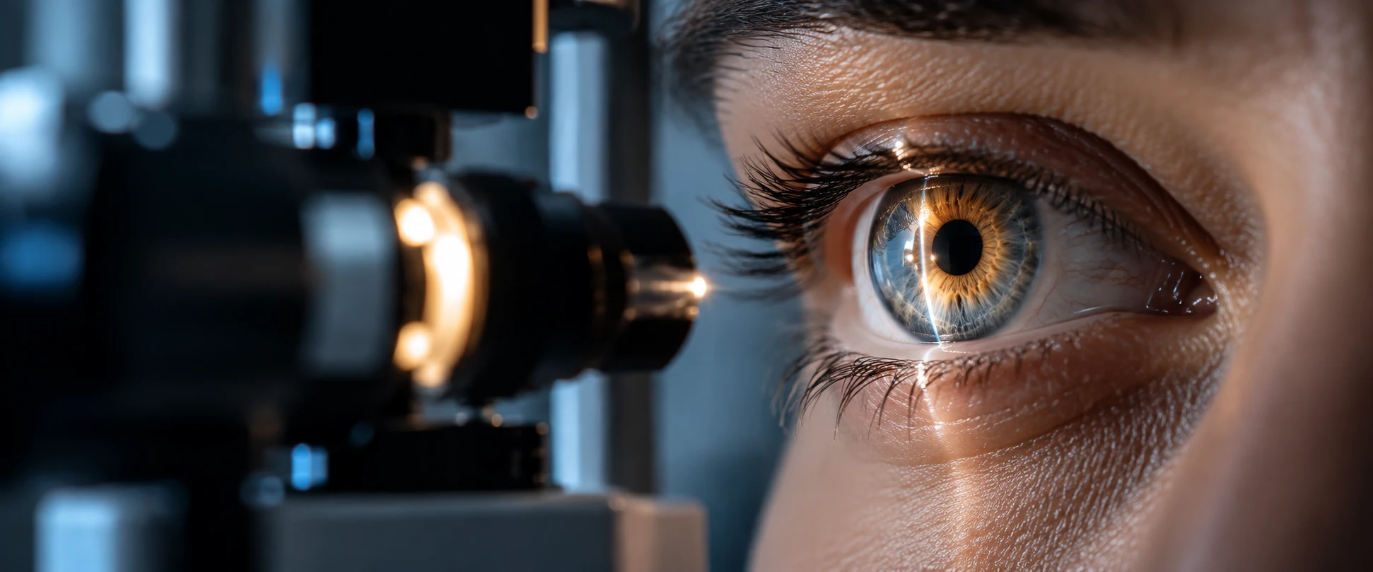
The approved hero is illustrative editorial artwork. It does not depict the study’s laboratory, a verified procedure or a treated participant. The study and this independently written explanation are distinct; study-derived findings and the graphic are attributed to Elia and colleagues under the paper’s Creative Commons Attribution 4.0 license. Creative Commons Attribution 4.0 license
FAQ
Frequently Asked Questions
Did this study restore sight in patients?
No. It studied patient-derived cells and retinal organoids in the laboratory, not treatment of people.
Is the experimental RP11 therapy approved?
This paper does not establish an approved RP11 treatment. Luxturna has a different FDA indication involving biallelic RPE65 mutations.
When will it be available?
The verified study provides no clinical availability date. Further in vivo safety and delivery work is needed.
PRESDA Dispatch
Stay Informed. Stay Aware.
A sharp briefing across AI, gaming, sport, business, world affairs, paparazzi, and lifestyle.